New directions in in vivo cell tracking, Dr. Paula Foster
Department of Physics and Astronomy
PHYSICS & ASTRONOMY COLLOQUIUM
via Zoom: Click here on 8 April 2021 @ 1:30
Dr. Paula Foster
Department of Medical Biophysics
Robarts Research Institute – Imaging
Western University
“New directions in in vivo cell tracking: Magnetic particle imaging”
ABSTRACT
Magnetic particle imaging (MPI) is an emerging, non-invasive imaging modality that directly detects the nonlinear magnetization response of superparamagnetic iron oxide (SPIO) tracers to an applied external magnetic field. SPIO particles have been used for over 20 years in magnetic resonance imaging (MRI), as cellular contrast agents to perform cell tracking. The MPI signal generated from SPIO is known to be specific, highly sensitive and linearly quantitative, making it well suited for in vivo tracking of cellular therapeutics.
MPI is built around a gradient magnet system. Two opposing electromagnets form a selection field, which is a strong gradient magnetic field that contains a field free region (FFR) near the isocenter where the magnetic field passes through zero. The selection field magnetically saturates the magnetization of all SPIOs except for those near the FFR, which experience no magnetic field. The FFR is rapidly rastered over the imaging volume, by changing the current through the electromagnets, to produce an image. When the FFR traverses a location containing SPIOs, the SPIOs magnetization changes and induces a voltage in a receiver coil. The voltages induced are linearly proportional to the number of SPIOs at the FFR location enabling quantification of SPIOs and estimates of cell number. This talk will focus on describing how MPI works, MPI sensitivity and resolution, tailored MPI tracers and specific applications for MPI related to cell tracking.
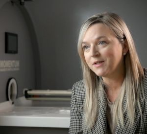
Canada’s first MPI system, (Momentum^TM, Magnetic Insight Inc. Alameda, CA), was commissioned at Western University in July 2019. Dr. Paula Foster is the Director of the MPI Laboratory at Western.

